Retinal Detachment Warning Signs to Look Out For

May 8, 2025
A close up of the inside of a human eye showing a retinal detachment

Read time: 5 minutes

Retinal detachment is a serious condition that can lead to vision loss if not treated quickly. It's important to know the warning signs so you can seek help right away. The retina is a thin layer of tissue at the back of the eye that helps us see. When the retina detaches, it pulls away from its normal position, which can cause permanent damage.

Imagine your eye is like a camera. The retina acts like the film or digital sensor that captures the images we see. If the retina is damaged or out of place, the camera can't take clear pictures. This is why recognizing the signs of retinal detachment early is so important. Quick treatment can make a big difference in saving your vision.

In this article, we will look at the key warning signs of retinal detachment, why it's urgent to seek treatment, and the medical solutions available to help protect your vision. Understanding these signs can help you and your loved ones keep your eyes healthy and your vision clear.

What Is Retinal Detachment?

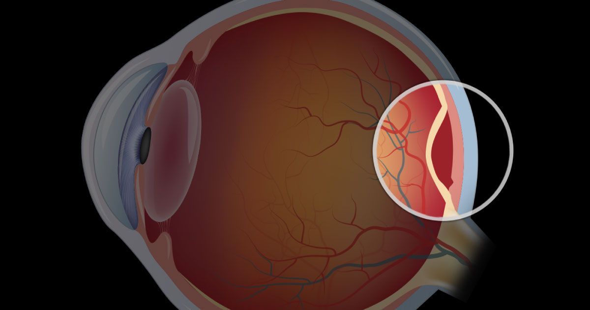
Retinal detachment happens when the retina, a thin layer of tissue in the back of the eye, separates from its normal position. The retina is crucial because it sends visual information to the brain. If it detaches, the retina can no longer function properly, leading to serious vision problems.

The eye is similar to a camera. The retina acts like the film or the digital sensor that captures the images we see. When the retina detaches, it's like removing the film from a camera, making it impossible to take a clear picture. Understanding the role of the retina helps highlight why quick action is essential if detachment occurs.

Key Warning Signs of Retinal Detachment

Recognizing the early signs of retinal detachment is vital to protect your vision. There are a few key symptoms to watch for:

  • Sudden Appearance of Floaters: These are tiny specks or strings that drift into your field of vision. They can be a sign of changes in the eye that might lead to retinal detachment.
  • Flashes of Light in Peripheral Vision: Seeing flashes of light, especially in the side vision, can indicate that the retina is under stress and pulling away from its normal position.
  • Shadow or Curtain Effect Over Vision: You might notice a dark shadow or curtain moving across your vision. This effect can start in one area and spread, indicating that the retina may be detaching.

These warning signs should never be ignored. If you experience any of these symptoms, seek medical attention immediately to prevent permanent vision loss.

To learn more about the types of sudden vision changes and their symptoms, read our article Decoding Sudden Vision Changes

The Urgency of Seeking Treatment

Seeking treatment for retinal detachment as soon as warning signs appear is crucial. Delay can lead to severe consequences, including permanent loss of vision. The longer the retina remains detached, the greater the risk of irreversible damage.

Delaying treatment can result in permanent vision loss, including blindness in the affected eye. Additionally, the longer the retina stays detached, the more complex and risky any potential surgery can become. Early intervention greatly reduces these complications and improves the likelihood of successful outcomes.

Importance of Early Detection for Preserving Vision

Early detection plays a vital role in the success of treatment. Immediate attention can significantly improve the chances of saving your vision. Identifying and addressing retinal detachment quickly helps prevent further complications and enhances the effectiveness of medical interventions.

What to Do If You Experience Symptoms

If you notice any warning signs such as floaters, flashes, or a shadow over your vision, it’s essential to contact an eye specialist immediately. Avoid delaying care or waiting to see if symptoms improve on their own. Prompt medical attention is critical for achieving the best possible outcome.

  • Medical Solutions and Treatments: There are several treatments available for retinal detachment, and the best approach depends on the severity and specific nature of the condition. Eye care professionals will assess the situation and recommend an appropriate treatment plan to preserve and restore vision.

  • Common Surgical Procedures: Scleral buckle surgery involves placing a silicone band around the eye to push the wall of the eye against the detached retina. Another common procedure is vitrectomy, where the vitreous gel pulling on the retina is removed and replaced with a gas bubble to help reattach the retina.

  • Laser Therapy Options: Laser photocoagulation uses a focused laser to create small burns around the retinal tear, helping to seal the retina back in place. Cryopexy is another method where a freezing probe is used to create scar tissue that secures the retina and prevents further detachment.

  • Recovery and Follow-Up Care: After surgery, following your doctor’s instructions carefully is essential for proper healing. Regular eye exams should be scheduled to monitor the health of your retina and catch any potential issues early. You may also need to follow temporary activity restrictions, such as avoiding heavy lifting or sudden movements, to protect your recovery and ensure optimal results.

The Takeaway

Retinal detachment is a serious condition that requires immediate attention. Understanding the key warning signs, such as sudden floaters, flashes of light, and a curtain effect over your vision, is crucial. Early detection and treatment can save your vision and prevent permanent damage.

If you experience any symptoms of retinal detachment, contact your local emergency eye care facility so you can be seen immediately.


If your eye injury or emergency occurs after hours and you are unable to reach an open facility, seek help at the nearest hospital emergency room, or call 911.


Always seek emergency care if you believe your eyesight is in jeopardy or if you are in severe pain.

Share this blog post on social or with a friend:

The information provided in this article is intended for general knowledge and educational purposes only and should not be construed as medical advice. It is strongly recommended to consult with an eye care professional for personalized recommendations and guidance regarding your individual needs and eye health concerns.


All of Urban Optiks Optometry's blog posts and articles contain information carefully curated from openly sourced materials available in the public domain. We strive to ensure the accuracy and relevance of the information provided. For a comprehensive understanding of our practices and to read our full disclosure statement, please click here.

OUR LATEST POSTS

The word fiction and fact are spelled out in wooden scrabble blocks on a rainbow background.
May 4, 2025
From vision "cures" to claims of corporate collusion, this article unpacks the most common eye care conspiracy theories and separates fact from fiction in modern optometry.
A surgeon is operating on a patient's eye in an operating room.
May 1, 2025
Read time: 4 minutes Vision correction surgery has undergone a remarkable transformation—from experimental corneal procedures to today’s precise, AI-assisted laser techniques. This journey reflects decades of scientific innovation, medical ingenuity, and an ever-growing understanding of the human eye. With each advancement, millions of people around the world have gained clearer, sharper vision and a better quality of life. Laying the Groundwork: Dr. Barraquer’s Early Vision The roots of modern refractive surgery trace back to the 1940s when Spanish ophthalmologist Dr. José Barraquer introduced the concept of altering the cornea’s shape to correct vision. Using a technique called keratomileusis, he manually removed and froze a section of the cornea, reshaping it before reimplanting it onto the eye. Though primitive by today’s standards, his foundational work laid the conceptual and surgical groundwork that future laser-based procedures would refine. The Excimer Laser Breakthrough A pivotal turning point arrived in the 1970s with the development of the excimer laser. Initially designed for industrial micro-machining, the laser’s ability to precisely remove organic material without causing surrounding thermal damage was a game-changer. In 1980, Dr. Rangaswamy Srinivasan demonstrated that the excimer laser could ablate human tissue with surgical precision—an insight that soon found profound applications in ophthalmology. PRK: The First FDA-Approved Laser Vision Correction By the late 1980s, these discoveries led to the development of Photorefractive Keratectomy (PRK)—a laser procedure that reshapes the cornea’s surface to correct refractive errors like myopia, hyperopia, and astigmatism. Approved by the FDA in 1995, PRK was the first laser eye surgery available in the United States and marked a significant shift from corrective lenses to permanent surgical solutions. While effective, PRK required longer healing times and post-operative discomfort due to the removal of the corneal epithelium. Nonetheless, it proved the efficacy of laser correction and paved the way for more advanced techniques. LASIK: A Game-Changer in Refractive Surgery Laser-Assisted In Situ Keratomileusis (LASIK) quickly followed. By creating a thin corneal flap before reshaping the underlying tissue with an excimer laser, LASIK dramatically improved patient comfort, accelerated healing, and reduced recovery time. It received FDA approval in 1998 and rapidly became the most popular refractive surgery worldwide. Over the years, LASIK has evolved through numerous enhancements: Bladeless LASIK: Uses femtosecond lasers for safer, more precise flap creation. Wavefront-guided LASIK: Tailors the laser ablation pattern to each eye’s unique optical imperfections, enhancing visual clarity and reducing side effects like halos or glare. Expanding Options: SMILE and Implantable Lenses Newer procedures now offer alternatives to traditional LASIK. One such innovation is SMILE (Small Incision Lenticule Extraction). This minimally invasive method removes a small lenticule of corneal tissue through a tiny incision - eliminating the need for a large flap and reducing the risk of dry eye or flap-related complications. For patients with thin corneas, extreme prescriptions, or dry eye syndrome, Implantable Collamer Lenses (ICL) or Refractive Lens Exchange (RLE) provide excellent non-laser-based options. ICL involves placing a flexible lens inside the eye, while RLE replaces the eye's natural lens, similar to cataract surgery but tailored for refractive correction. Learn more about SMILE here. What’s Next: AI, Smart Lenses, and Personalized Eye Care As technology advances, the future of vision correction is becoming even more personalized, efficient, and precise. Current research and emerging innovations include: AI-Powered Diagnostics: Machine learning algorithms analyze corneal shape, thickness, and wavefront data to optimize surgical planning. Next-Generation IOLs: Intraocular lenses now offer multifocal, extended depth-of-focus, and light-adjustable features for better post-cataract outcomes. Minimally Invasive Procedures: Smaller incisions and non-invasive techniques are reducing downtime and surgical risk. Presbyopia Solutions: Using preserved corneal lenticules from SMILE to potentially reverse age-related reading vision loss. Enhanced Wavefront & Topography Tools: Improved mapping systems enable ultra-customized ablation profiles. AI-Assisted Surgery Platforms: Real-time intraoperative guidance tools help surgeons make micro-adjustments for superior results. The Takeaway What began as a bold concept in a 1940s laboratory has blossomed into a diverse, high-tech field with solutions for nearly every refractive need. Today’s surgical options are faster, safer, and more tailored than ever before, and the horizon continues to expand with each innovation. As artificial intelligence, biomechanics, and surgical precision continue to evolve, so too will the ways we restore vision - bringing clarity to life in ways once thought impossible.
A woman wearing oversized aviator glasses and a leather jacket is standing on a set of stairs.
April 28, 2025
90s eyewear styles are back! From iconic frames to modern looks, discover how to wear them and where to find the best picks.
A group of business people wearing glasses are standing next to each other in an office.
April 24, 2025
Discover why eyewear is essential for business. Learn how it boosts confidence, enhances professionalism, and offers practical benefits for your career.
A close up of a person 's eye with a reflection of small rays of light
April 21, 2025
Discover the biomechanics of eye movement—how muscles, nerves, and the brain work together for precise vision. Learn about saccades, tracking, and reflexes.
A close up of a person 's eye with a robotic illustration overlay.
April 16, 2025
Explore how nanotechnology is revolutionizing lens manufacturing - from smart contact lenses that monitor health to advanced eyewear coatings.
A woman in a red hat is painting a mural with eyes on a wall.
April 14, 2025
Explore how vision symbolizes knowledge, truth, and perception in art and literature, from ancient myths to modern technology, shaping cultural understanding.
A woman wearing sunglasses with grey gradient lenses is looking up.
April 11, 2025
Discover the stylish appeal and practical benefits of gradient lenses. Learn how to choose and care for gradient lenses for ultimate eye comfort.
A pair of vintage glasses laying on top of an open newspaper
April 7, 2025
Explore the history of vision correction, from Egyptian magnifiers to medieval reading stones, and how ancient innovations shaped modern optometry.
A man is sitting at a table with a tray of herbs on it.
April 3, 2025
Discover TCM and Ayurveda’s natural eye care with herbs, diet, and holistic practices like acupuncture and Triphala for better vision and overall well-being.
The word fiction and fact are spelled out in wooden scrabble blocks on a rainbow background.
May 4, 2025
From vision "cures" to claims of corporate collusion, this article unpacks the most common eye care conspiracy theories and separates fact from fiction in modern optometry.
A surgeon is operating on a patient's eye in an operating room.
May 1, 2025
Read time: 4 minutes Vision correction surgery has undergone a remarkable transformation—from experimental corneal procedures to today’s precise, AI-assisted laser techniques. This journey reflects decades of scientific innovation, medical ingenuity, and an ever-growing understanding of the human eye. With each advancement, millions of people around the world have gained clearer, sharper vision and a better quality of life. Laying the Groundwork: Dr. Barraquer’s Early Vision The roots of modern refractive surgery trace back to the 1940s when Spanish ophthalmologist Dr. José Barraquer introduced the concept of altering the cornea’s shape to correct vision. Using a technique called keratomileusis, he manually removed and froze a section of the cornea, reshaping it before reimplanting it onto the eye. Though primitive by today’s standards, his foundational work laid the conceptual and surgical groundwork that future laser-based procedures would refine. The Excimer Laser Breakthrough A pivotal turning point arrived in the 1970s with the development of the excimer laser. Initially designed for industrial micro-machining, the laser’s ability to precisely remove organic material without causing surrounding thermal damage was a game-changer. In 1980, Dr. Rangaswamy Srinivasan demonstrated that the excimer laser could ablate human tissue with surgical precision—an insight that soon found profound applications in ophthalmology. PRK: The First FDA-Approved Laser Vision Correction By the late 1980s, these discoveries led to the development of Photorefractive Keratectomy (PRK)—a laser procedure that reshapes the cornea’s surface to correct refractive errors like myopia, hyperopia, and astigmatism. Approved by the FDA in 1995, PRK was the first laser eye surgery available in the United States and marked a significant shift from corrective lenses to permanent surgical solutions. While effective, PRK required longer healing times and post-operative discomfort due to the removal of the corneal epithelium. Nonetheless, it proved the efficacy of laser correction and paved the way for more advanced techniques. LASIK: A Game-Changer in Refractive Surgery Laser-Assisted In Situ Keratomileusis (LASIK) quickly followed. By creating a thin corneal flap before reshaping the underlying tissue with an excimer laser, LASIK dramatically improved patient comfort, accelerated healing, and reduced recovery time. It received FDA approval in 1998 and rapidly became the most popular refractive surgery worldwide. Over the years, LASIK has evolved through numerous enhancements: Bladeless LASIK: Uses femtosecond lasers for safer, more precise flap creation. Wavefront-guided LASIK: Tailors the laser ablation pattern to each eye’s unique optical imperfections, enhancing visual clarity and reducing side effects like halos or glare. Expanding Options: SMILE and Implantable Lenses Newer procedures now offer alternatives to traditional LASIK. One such innovation is SMILE (Small Incision Lenticule Extraction). This minimally invasive method removes a small lenticule of corneal tissue through a tiny incision - eliminating the need for a large flap and reducing the risk of dry eye or flap-related complications. For patients with thin corneas, extreme prescriptions, or dry eye syndrome, Implantable Collamer Lenses (ICL) or Refractive Lens Exchange (RLE) provide excellent non-laser-based options. ICL involves placing a flexible lens inside the eye, while RLE replaces the eye's natural lens, similar to cataract surgery but tailored for refractive correction. Learn more about SMILE here. What’s Next: AI, Smart Lenses, and Personalized Eye Care As technology advances, the future of vision correction is becoming even more personalized, efficient, and precise. Current research and emerging innovations include: AI-Powered Diagnostics: Machine learning algorithms analyze corneal shape, thickness, and wavefront data to optimize surgical planning. Next-Generation IOLs: Intraocular lenses now offer multifocal, extended depth-of-focus, and light-adjustable features for better post-cataract outcomes. Minimally Invasive Procedures: Smaller incisions and non-invasive techniques are reducing downtime and surgical risk. Presbyopia Solutions: Using preserved corneal lenticules from SMILE to potentially reverse age-related reading vision loss. Enhanced Wavefront & Topography Tools: Improved mapping systems enable ultra-customized ablation profiles. AI-Assisted Surgery Platforms: Real-time intraoperative guidance tools help surgeons make micro-adjustments for superior results. The Takeaway What began as a bold concept in a 1940s laboratory has blossomed into a diverse, high-tech field with solutions for nearly every refractive need. Today’s surgical options are faster, safer, and more tailored than ever before, and the horizon continues to expand with each innovation. As artificial intelligence, biomechanics, and surgical precision continue to evolve, so too will the ways we restore vision - bringing clarity to life in ways once thought impossible.
A woman wearing oversized aviator glasses and a leather jacket is standing on a set of stairs.
April 28, 2025
90s eyewear styles are back! From iconic frames to modern looks, discover how to wear them and where to find the best picks.
A group of business people wearing glasses are standing next to each other in an office.
April 24, 2025
Discover why eyewear is essential for business. Learn how it boosts confidence, enhances professionalism, and offers practical benefits for your career.
A close up of a person 's eye with a reflection of small rays of light
April 21, 2025
Discover the biomechanics of eye movement—how muscles, nerves, and the brain work together for precise vision. Learn about saccades, tracking, and reflexes.