Alzheimer’s, Dementia and the Eye: What Research Shows
Read time: 6 minutes
What if changes in your eyes could someday help doctors understand what is happening in your brain? That possibility is driving an exciting area of Alzheimer's and dementia research. Scientists are discovering that the retina—the light-sensitive tissue at the back of the eye—may contain clues about changes occurring in the brain, sometimes before noticeable symptoms develop.
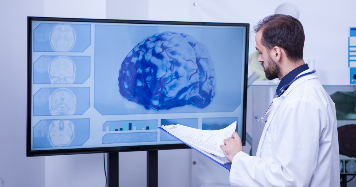
The retina is actually part of the central nervous system and shares important characteristics with brain tissue, including nerve cells and blood vessels. Researchers are studying whether advanced retinal imaging could eventually help identify or monitor changes associated with Alzheimer's disease.
An important distinction: A routine eye examination or retinal scan cannot currently diagnose Alzheimer's disease or dementia. The research is promising, but these technologies are not yet established as clinical screening tests.
Alzheimer's Disease and Dementia: What's the Difference?
Although the terms are often used interchangeably, they aren't the same thing. Dementia describes a decline in memory, reasoning, thinking, or other cognitive abilities significant enough to interfere with daily life. Alzheimer's disease is the most common cause of dementia, but other forms include:
- Vascular dementia
- Lewy body dementia
- Frontotemporal dementia
- Mixed dementia involving multiple disease processes
Different types of dementia affect the brain in different ways, which may also influence vision and visual processing. Understanding these differences is important because not all cognitive or visual changes have the same underlying cause.
Why Scientists Are Studying the Retina
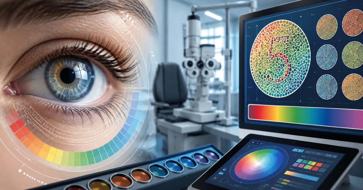
The retina offers researchers something unusual: nervous-system tissue that can be examined directly without invasive procedures. Modern technologies such as optical coherence tomography (OCT) produce highly detailed images of retinal layers, while OCT angiography (OCT-A) can map tiny retinal blood vessels without injecting dye.
Researchers have identified several retinal differences in some people with Alzheimer's disease or mild cognitive impairment, including:
- Thinning of retinal nerve fiber and ganglion cell layers
- Changes in macular thickness
- Reduced retinal blood-vessel density
- Differences in retinal circulation and vascular structure
These findings are significant because Alzheimer's involves nerve-cell damage, inflammation, abnormal protein accumulation, and vascular changes in the brain. Scientists are investigating whether similar processes leave detectable signs in the retina and whether those changes could eventually become useful indicators of neurological health.
Learn more about how Urban Optiks Optometry uses detailed retinal imaging to evaluate the retina, optic nerve, macula, and retinal blood vessels.
What Have Researchers Actually Discovered?
Two proteins closely associated with Alzheimer's disease are amyloid-beta, which forms plaques between nerve cells, and abnormal tau proteins, which accumulate inside nerve cells. Studies examining donated retinal tissue have identified amyloid deposits, inflammation, vascular abnormalities, and changes in nerve tissue among some people with Alzheimer's disease.
A study supported by the National Institute on Aging found higher amounts of retinal amyloid in people with Alzheimer's disease and mild cognitive impairment compared with cognitively unimpaired individuals. Researchers also observed associations between retinal findings and the severity of changes in the brain. These discoveries suggest that the retina could eventually provide another way to study neurodegenerative disease.
However, this does not mean an optometrist can currently examine your retina and determine whether you have Alzheimer's. Researchers still need to establish which findings are sufficiently accurate, consistent, and specific for clinical use.
Can an OCT Scan Detect Alzheimer's?
Not by itself. OCT is already an important diagnostic tool for examining retinal and optic nerve health, particularly for conditions such as glaucoma and retinal disease. Research has identified changes in retinal thickness and structure among some patients with Alzheimer's disease, and a 2026 systematic review examined their potential as early biomarkers.
The challenge is that similar retinal changes can occur with normal aging, glaucoma, diabetes, high myopia, and other eye conditions. This makes it difficult to distinguish changes associated with Alzheimer's from unrelated conditions. Researchers are also studying OCT angiography because Alzheimer's can affect blood vessels in the brain, and some studies have found reduced retinal vascular density in people with Alzheimer's or mild cognitive impairment.
Although these findings are encouraging, neither OCT nor OCT angiography is currently a proven stand-alone screening test for Alzheimer's disease. Additional research is needed before retinal imaging can reliably distinguish Alzheimer's-related changes from other common eye conditions.
A closer look at retinal imaging: A 2026 systematic review examined OCT findings being studied as possible retinal biomarkers for early Alzheimer’s disease.
Dementia Can Affect Vision Even When the Eyes Are Healthy
One of the most important connections between dementia and vision has less to do with the eyes themselves and more to do with how the brain interprets visual information. Your eyes collect visual information, but your brain must process and interpret it. When dementia affects regions responsible for visual processing, everyday activities can become more difficult even when the eyes are otherwise healthy.
Someone experiencing cognitive decline may have difficulty with:
- Depth perception: Judging distances, navigating stairs, or reaching for objects
- Contrast sensitivity: Distinguishing objects from similarly colored backgrounds
- Spatial awareness: Navigating familiar spaces or recognizing where objects are located
- Visual recognition: Identifying objects, faces, or complex scenes
- Reading and interpretation: Processing written information or understanding visual surroundings
These difficulties can sometimes be mistaken for ordinary poor eyesight. The Alzheimer's Association identifies changes in visual images and spatial relationships as possible warning signs of Alzheimer's disease, although similar symptoms can also occur with other eye and neurological conditions.
Learn about the warning signs of Alzheimer's disease
How Changes in Vision Affect Everyday Life
Visual acuity - how well someone reads letters on an eye chart—is only one part of healthy vision. Contrast sensitivity, depth perception, and visual processing are equally important for safely navigating the world. Someone may be able to read relatively small letters during an eye examination yet struggle to interpret objects and surroundings in everyday situations.
For someone experiencing dementia-related visual difficulties, ordinary environments may become unexpectedly challenging:
- A white plate may be difficult to distinguish from a white tablecloth.
- A pale stair may blend into the surrounding flooring.
- A doorway or furniture edge may be harder to judge.
- Complex patterns or cluttered spaces may create confusion.
- Driving may become difficult because of changes in distance judgment or spatial awareness.
These symptoms don't necessarily indicate dementia. Cataracts, glaucoma, retinal disease, outdated prescriptions, and other vision conditions can produce similar difficulties. That distinction is one reason comprehensive eye examinations remain important, particularly when someone is experiencing changes in memory or cognitive function.
Could Artificial Intelligence Help Detect Alzheimer's Earlier?
Artificial intelligence is becoming another important part of retinal research. Scientists are developing AI systems that analyze retinal photographs, OCT scans, and vascular imaging for subtle patterns involving nerve tissue, blood vessels, and retinal structure that may be difficult to recognize individually.
A 2026 review examining 63 studies of AI-based retinal imaging for Alzheimer's detection found encouraging potential, but also emphasized the need for stronger clinical validation. Before these systems can become dependable screening tools, researchers must demonstrate that they work accurately across large, diverse populations and can distinguish Alzheimer's-related findings from normal aging and unrelated eye disease.
The opportunity is exciting, but AI-based retinal screening for Alzheimer's is still experimental. It may eventually complement established neurological testing, but it is not currently a substitute for a comprehensive medical evaluation.
Why Regular Eye Exams Still Matter for People With Dementia
Although eye examinations cannot currently diagnose dementia, maintaining healthy vision can make a meaningful difference for people experiencing cognitive decline. Common vision problems—including cataracts, dry eye, glaucoma, macular degeneration, and outdated eyeglass prescriptions—can make daily activities more challenging when combined with difficulties in memory or visual processing.
Identifying and treating correctable eye conditions may help people make better use of their remaining visual abilities. Clearer vision can also make it easier to recognize familiar surroundings, navigate daily activities, and maintain independence.
Small Changes at Home Can Also Help
For someone experiencing dementia-related visual difficulties, a few practical adjustments may improve comfort and navigation:
- Increase lighting while minimizing harsh glare.
- Use contrasting colors to distinguish objects from backgrounds.
- Keep walkways clear and furniture in familiar locations.
- Make stair edges easier to identify.
- Use larger, easy-to-read labels.
- Reduce visually confusing patterns and clutter.
These adjustments do not treat dementia, but they may help make everyday environments easier to understand and navigate. New or worsening vision problems should never automatically be attributed to dementia, particularly when symptoms appear suddenly or interfere significantly with daily activities. Sudden vision changes require prompt medical evaluation.
The Takeaway
The connection between the eyes and brain is real, and research into retinal biomarkers for Alzheimer's disease is advancing. Scientists have identified structural, vascular, inflammatory, and molecular differences in the retinas of some people with Alzheimer's disease and mild cognitive impairment. Technologies such as OCT, OCT angiography, and artificial intelligence may eventually provide additional tools for identifying or monitoring neurodegenerative disease.
For now, however, a routine eye exam or retinal scan cannot reliably diagnose Alzheimer's disease or dementia. Larger, standardized studies are needed before these technologies can become part of routine neurological screening.
What comprehensive eye care can already do is identify and manage vision problems that may complicate everyday life, particularly for people experiencing cognitive decline. At Urban Optiks Optometry, comprehensive eye examinations and advanced retinal imaging help evaluate the health of the retina, optic nerve, and other important eye structures. When visual changes occur alongside memory, reasoning, or navigation difficulties, coordinated evaluation with a primary-care physician or neurological specialist may also be appropriate.
Share this blog post on social or with a friend:
The information provided in this article is intended for general knowledge and educational purposes only and should not be construed as medical advice. It is strongly recommended to consult with an eye care professional for personalized recommendations and guidance regarding your individual needs and eye health concerns.
All of Urban Optiks Optometry's blog posts and articles contain information carefully curated from openly sourced materials available in the public domain. We strive to ensure the accuracy and relevance of the information provided. For a comprehensive understanding of our practices and to read our full disclosure statement, please click here.