Comprehensive Eye Exam
At Urban Optiks Optometry, our state-of-the-art eye examinations use the latest equipment to ensure an accurate and comfortable prescription. Computerized technology is incorporated into the autorefractor, auto lensometer, peripheral visual field and glaucoma testing for a comprehensive evaluation of your overall eye health. Our doctors work patiently with you to ensure that your prescription best suits your vision requirements whether it be for glasses or contact lenses.
What is a Comprehensive Eye Exam?
During a comprehensive eye exam, your eye doctor does more than just determine your glasses and contacts prescription. Only a small percentage of your time during an exam is actually spent determining your prescription.
The majority of the testing time is devoted to tests such as visual field, tonometry, retinal evaluation, and dry eye screenings. Your optometrist is often the first line of defense in the early detection of chronic diseases such as high blood pressure and diabetes. As many eye and vision problems have no obvious signs or symptoms, you may be completely unaware of them. These tests are just a few of the ways that optometrists can check your overall health and well-being.
Even if you can pass a screening and see 20/20, you should still have annual eye examinations to check the health of your eyes. Simply put… EVERYONE, EVERY YEAR.
Here's what to expect during your eye exam
Our optometrists use a variety of tests and procedures to examine your eyes, ranging from simple to complex. From having you read an eye chart to using a high-powered retinal camera to visualize the tiny structures inside of your eyes, all of these tests are important to the process of accurately evaluating the health of your eyes and recommending an accurate prescription. In order to fully evaluate your vision and the health of your eyes, you should expect to spend about an hour for your examination.
Below are some of the tests that you are likely to participate in during a routine comprehensive eye exam.
Comprehensive Eye Exam Testing
Visual Field Analyzer
In order to check for the possible presence of blind spots (scotomas) in your peripheral vision, our optometrists will perform a visual field test. He’ll ask you to place your forehead against the visual field analyzer device and to hold a button in your hand. You will see a white screen inside with a black dot at the center. While focusing on the black dot, you will see a series of squiggly lines pop up in different quadrants of the white screen. Each time you see one of these squiggly lines, you will click the button in your hand. Blind spots in your vision can help the doctor to diagnose important health issues.
Visual Acuity Test
A visual acuity test, which measures the sharpness of your vision, is often one of the first tests performed during your eye exam. To measure your distance visual acuity, the doctor may ask you what the smallest line you can read is on an eye chart projected on a screen, mirror or TV. A similar test is performed for your near vision, typically using a small, hand-held acuity chart.
Normal visual acuity is written as 20/20. If you have reduced vision, it will be recorded as less than 20/20, such as 20/400. If you have 20/400 vision, it means that an object that you can see from 20 feet away, a normal 20/20 sighted person can see from 400 feet away. There are many ways to check how your eyes work together, however, the cover test is the simplest and most common.
During a cover test, you focus on a small object across the room and the doctor will cover each of your eyes one at a time while you stare at the target. While doing this, they’ll assess whether the uncovered eye must move to pick up the fixation target, which could indicate vision problems such as strabismus or amblyopia.
Glaucoma Test
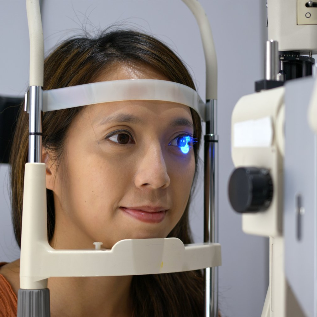
The glaucoma test, a.k.a. the “puff-of-air” test, technically known as non-contact tonometry, or NCT, is designed to measure the pressure inside of your eye and is often perceived to be the most difficult part of your eye exam. Once again you will be asked to put your chin on the machine’s chin rest, this time looking at a light inside the machine. The machine will puff a small burst of air at your open eye and based on your eye’s resistance to the puff of air will produce a number that indicates your intraocular pressure (IOP). It is completely painless, and the tonometer does not touch your eye. If your IOP number is high, this is an indicator that you could be at risk for glaucoma. Other than an elevated IOP, there are no warning signs of glaucoma until you start to have vision loss. This is just one reason that routine eye exams are essential to protecting your eyesight and overall health.
Autorefractor Keratometer
The doctor will use an autorefractor as a starting point in determining your prescription. With this piece of equipment, you will be asked to rest your face in a chin rest and focus on a distant image inside of the machine. An autorefractor determines the lens power required by your eye to accurately focus light on your retina, giving the doctor a baseline prescription in an accurate & time-saving manner. This is especially useful in certain cases where a patient cannot sit still, pay attention, or interact with the eye doctor adequately for the amount of time necessary for a manual refraction. The autorefraction machine can produce an accurate baseline prescription in only a few seconds!
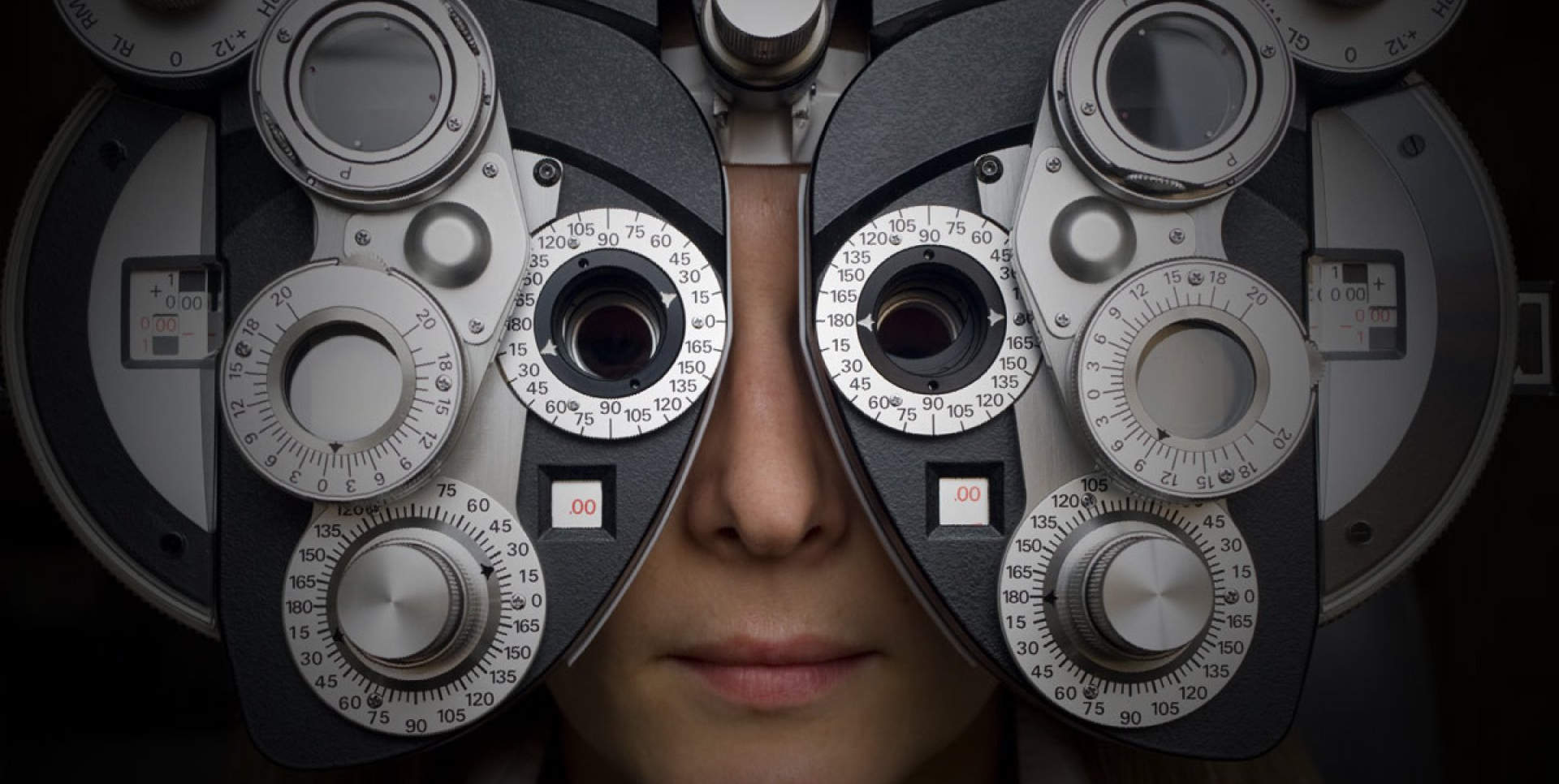
Digital Refraction
This is the test used to determine your exact eyeglass prescription. During a manual refraction, the doctor will put an instrument called a phoropter in front of your eyes. You will be asked to view an eye chart or another testing image through a series of lenses. You will be asked about how you perceive the images through each lens. Based on your answers, the doctor will be able to fine-tune the lens power until a final prescription is reached. The refraction determines your prescription for whichever accommodation disorder you may be experiencing: hyperopia (farsightedness), myopia (nearsightedness), astigmatism or presbyopia.
Slit-lamp Biomicroscopy Examination
When it comes time to examine the health of your eyes, the doctor typically uses a digital slit lamp, also known as a biomicroscope, to get a highly magnified view of the structures of your eye, allowing them to detect any signs of infection or disease. While you have your chin on the chin rest of the slit lamp, they will shine a light at your eye. The doctor will look through a set of oculars (similar to a microscope) and examine each part of your eye, starting with the front of your eye (conjunctiva, lids, iris, cornea). In some cases, they’ll also view the inside of your eye (optic nerve, retina, macula) with the slit lamp. However, most doctors prefer to perform a dilation or retinal photography (see below) to get an even better view of the inside of your eye than the slit-lamp can provide. Optometrists are often the first to detect a wide range of eye conditions with use of the slit-lamp examination, including macular degeneration, cataracts, corneal ulcers, diabetic retinopathy, and more.
Pupil Dilation
To obtain a better view of your eye’s internal structures, the doctor may need to enlarge your pupils with the use of dilating drops. The drops usually take about 20 to 30 minutes to start working. Once they are dilated, the doctor will use a variety of instruments to look through your enlarged pupil. As a result of pupil dilation, you may experience sensitivity to light and reduced visual clarity at near which usually lasts for 4-6 hours afterward. It is recommended that you bring sunglasses to wear afterward and don’t plan on doing any up-close work after your dilation.
Digital Retinal Imaging & OCT
In lieu of dilation, you may have the option of having the inside of your eyes viewed using a digital retinal imaging device. Using a high-powered "camera" lens, the equipment captures an image of the inside of your eye. This photo is then loaded into your electronic medical records and serves as a baseline record of the health of your eyes. At future examinations, subsequent photographs will allow the doctor to compare the images to see how the internal structures of your eyes are changing.
Most insurance plans do not cover retinal photography as part of a comprehensive eye examination, therefore, you may be charged an additional fee for this test.
Color Blindness Test
If you suspect that you may have a problem with viewing colors accurately, a color blindness test can be performed. In addition to detecting color vision deficiencies, color blindness tests can alert the doctor to possible eye health problems that may affect your color vision.
Other Eye Tests
In some cases, the doctor may recommend other, more specialized eye tests.