Hypertensive Retinopathy vs. Diabetic Retinopathy

November 29, 2023
Person with bloodshot eyes

Read time: 6 minutes


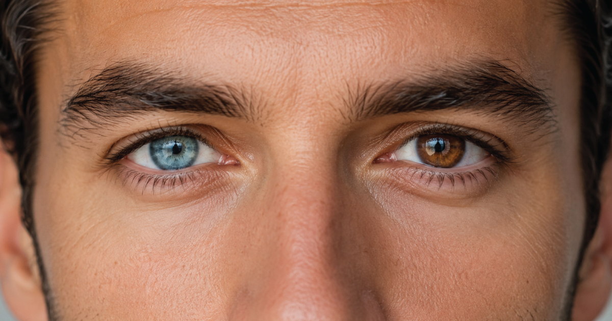
The human eye, with its intricate network of blood vessels, is a complex organ that can be profoundly affected by systemic diseases. Two conditions that specifically target the delicate blood vessels within the eye are hypertensive retinopathy and diabetic retinopathy.


In this blog post, we will delve into the historical and contemporary aspects of these conditions, exploring their causes, symptoms, and treatment options. In addition, understanding when these conditions typically begin can aid in proactive health management. 


Understanding Retinopathy


The understanding of retinopathy has evolved over centuries, paralleling advancements in medical science. The earliest observations of retinal disorders can be traced back to ancient civilizations, where limited anatomical knowledge hindered a comprehensive understanding. It wasn't until the 19th century that detailed observations of the eye and its pathology began to emerge.


One of the pioneers in retinal research was the German physician Hermann von Helmholtz, who invented the ophthalmoscope in 1851. This revolutionary device allowed for the direct visualization of the retina, paving the way for a more accurate diagnosis of retinal disorders. As medical knowledge advanced, so did the recognition of hypertensive and diabetic retinopathy as distinct entities.


Typical Onset Age of Hypertensive Retinopathy


Hypertensive retinopathy tends to develop in adults, typically after years of uncontrolled high blood pressure. While there isn't a specific age range for its onset, it is more commonly observed in middle-aged and older individuals.


The risk of hypertensive retinopathy increases with age, especially in those with prolonged periods of untreated high blood pressure. Regular blood pressure monitoring becomes crucial as individuals advance in age.


Typical Onset Age of Diabetic Retinopathy


Diabetic retinopathy is closely linked to the duration of diabetes rather than a specific age. It can, however, start to manifest after several years of living with diabetes. Both type 1 and type 2 diabetes can lead to diabetic retinopathy.


The longer an individual has diabetes, the higher the risk of developing diabetic retinopathy. Onset may occur earlier in life for those diagnosed with diabetes at a young age.


While hypertensive retinopathy may be more prevalent in middle-aged and older adults due to prolonged exposure to high blood pressure, diabetic retinopathy is closely tied to the duration of diabetes rather than a specific age. Regular health check-ups, blood pressure monitoring, and diabetes management play crucial roles in mitigating the risk and progression of these retinopathies across different age groups.


Causes and Characteristics


Hypertensive Retinopathy


  • Causes: Hypertensive retinopathy is a manifestation of long-term high blood pressure affecting the blood vessels in the retina. The increased pressure forces the blood vessels to narrow and become more rigid over time. This vascular damage can lead to a variety of changes in the retina, affecting both the smaller arterioles and the larger vessels.


  • Clinical Features: The hallmark signs of hypertensive retinopathy include arteriolar narrowing, arteriovenous nicking, and flame-shaped hemorrhages. As the condition progresses, more severe manifestations such as cotton-wool spots and optic disc edema may occur. Cotton-wool spots represent areas of nerve fiber layer infarction, a consequence of compromised blood supply to the retinal nerve fibers.


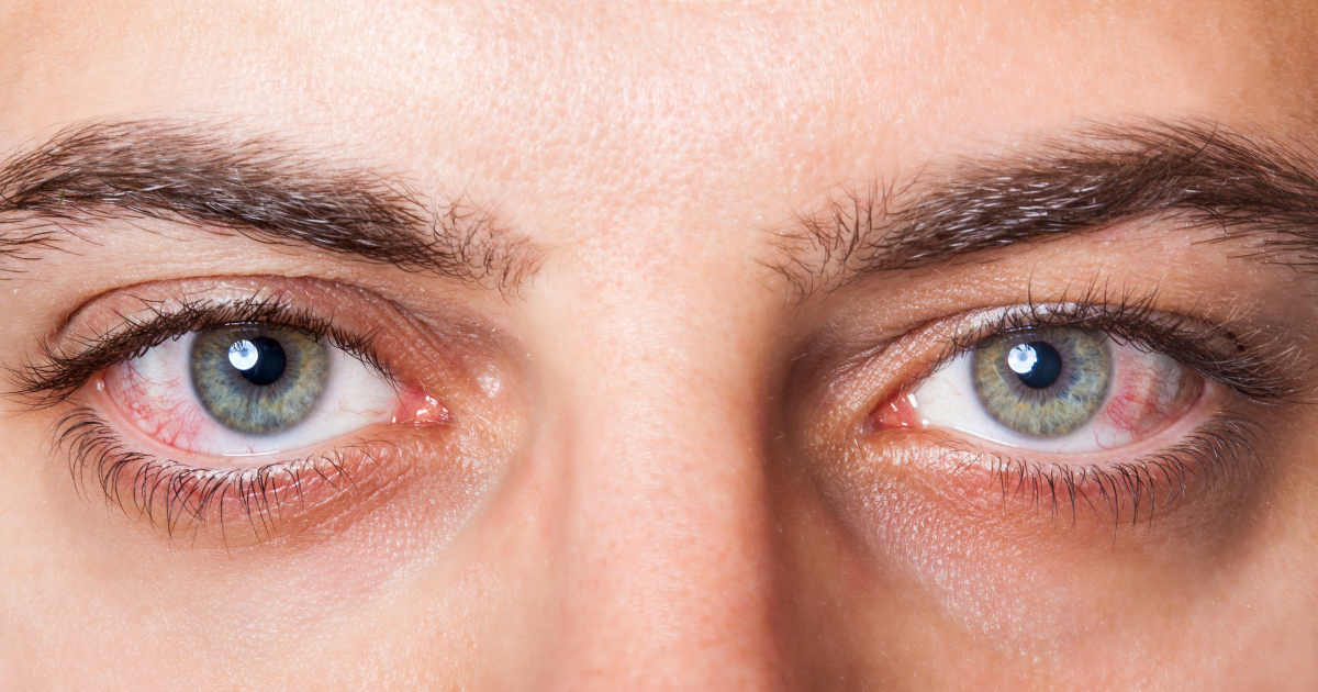
  • Symptoms: In the early stages, hypertensive retinopathy may be asymptomatic. However, as the disease advances, symptoms such as blurred vision, headaches, and visual disturbances may manifest. Regular eye examinations are crucial for early detection and management.


Diabetic Retinopathy


  • Causes: Diabetic retinopathy, as the name suggests, is a complication of diabetes mellitus. Prolonged exposure to high blood sugar levels can damage the small blood vessels in the retina, leading to a cascade of pathological changes. Both type 1 and type 2 diabetes patients are at risk, emphasizing the importance of glycemic control.


  • Clinical Features: The progression of diabetic retinopathy is categorized into non-proliferative and proliferative stages. Non-proliferative diabetic retinopathy (NPDR) is characterized by microaneurysms, hemorrhages, and lipid exudates. As the disease advances to the proliferative stage, the growth of abnormal blood vessels on the retina's surface becomes a concern, posing a higher risk of bleeding into the vitreous, retinal detachment, and severe vision loss.


  • Symptoms: Similar to hypertensive retinopathy, diabetic retinopathy may initially be asymptomatic. As the condition progresses, patients may experience symptoms such as floaters, blurred vision, and difficulty perceiving colors. Routine eye examinations are critical for early detection and intervention.


Diagnosis and Evaluation


Both hypertensive and diabetic retinopathy are diagnosed through a comprehensive eye examination, which includes visual acuity testing, dilated fundus examination, and imaging studies such as fluorescein angiography and optical coherence tomography (OCT).


  • Fluorescein Angiography: This diagnostic procedure involves the intravenous injection of a fluorescent dye, which highlights the retinal blood vessels. It aids in identifying areas of ischemia, leakage, and abnormal blood vessel growth.


  • Optical Coherence Tomography (OCT): OCT is a non-invasive imaging technique that provides detailed cross-sectional images of the retina. It is particularly useful in assessing the thickness of the retinal layers and detecting the presence of macular edema.


Treatment Approaches


The management of hypertensive and diabetic retinopathy involves a combination of lifestyle modifications, medical therapy, and, in some cases, surgical interventions.


Hypertensive Retinopathy


  • Blood Pressure Control: The primary focus in hypertensive retinopathy management is controlling blood pressure. Lifestyle modifications, including a low-sodium diet, regular exercise, and medication adherence, play a crucial role in achieving optimal blood pressure levels.
  • Anti-Hypertensive Medications: Medications such as angiotensin-converting enzyme (ACE) inhibitors and angiotensin II receptor blockers (ARBs) are commonly prescribed to manage hypertension and mitigate its impact on the retinal vasculature.
  • Regular Monitoring: Close monitoring of blood pressure and regular eye examinations are essential components of managing hypertensive retinopathy. Timely intervention can prevent progression to more severe stages of the disease.


Diabetic Retinopathy


  • Glycemic Control: Achieving and maintaining tight glycemic control is fundamental in preventing and managing diabetic retinopathy. Diabetic patients should work closely with their healthcare team to monitor and manage blood sugar levels effectively.
  • Intravitreal Injections: In cases of diabetic macular edema or proliferative diabetic retinopathy, intravitreal injections of anti-vascular endothelial growth factor (VEGF) medications or corticosteroids may be recommended to reduce swelling and inhibit abnormal blood vessel growth.
  • Laser Photocoagulation: Laser therapy is often used to treat specific complications of diabetic retinopathy, such as macular edema and proliferative disease. The laser helps to seal leaking blood vessels and reduce the risk of further vision loss.
  • Vitrectomy: In advanced cases with significant vitreous hemorrhage or tractional retinal detachment, vitrectomy surgery may be considered. During this procedure, the vitreous gel is removed and replaced with a clear solution.


Prognosis and Prevention


Both hypertensive and diabetic retinopathy require ongoing management and monitoring to prevent complications and preserve vision. When effectively managed, the prognosis for hypertensive retinopathy is generally favorable. However, untreated or poorly controlled hypertension can lead to progressive retinal damage and, in severe cases, permanent vision loss.


The prognosis for diabetic retinopathy varies based on the stage of the disease at diagnosis and the effectiveness of management strategies. Early detection and intervention significantly improve outcomes, emphasizing the importance of regular eye examinations for individuals with diabetes.


The Takeaway


As we've learned, hypertensive retinopathy and diabetic retinopathy are distinct yet interconnected conditions that highlight the intricate relationship between systemic health and ocular well-being. Historical advancements in ophthalmic research and modern diagnostic tools have significantly improved our ability to detect and manage these conditions effectively.


As we continue to refine our understanding of the pathophysiology of these retinopathies, ongoing research may unveil novel therapeutic approaches, providing hope for improved outcomes and a brighter future for individuals at risk of vision-threatening complications. Regular eye examinations at award-winning Urban Optiks Optometry, early intervention, and collaborative care between healthcare professionals and patients remain the cornerstone of preserving vision in the face of these potentially devastating conditions. Schedule your annual eye exam today!

Share this blog post on social or with a friend:

The information provided in this article is intended for general knowledge and educational purposes only and should not be construed as medical advice. It is strongly recommended to consult with an eye care professional for personalized recommendations and guidance regarding your individual needs and eye health concerns.


All of Urban Optiks Optometry's blog posts and articles contain information carefully curated from openly sourced materials available in the public domain. We strive to ensure the accuracy and relevance of the information provided. For a comprehensive understanding of our practices and to read our full disclosure statement, please click here.

OUR LATEST POSTS

Woman wearing yellow-rimmed sunglasses and a cream-colored top with arms above her head.
October 22, 2025
Discover why some designs endure in fashion. Explore how eyewear luxury brands like Urban Optiks Optometry blend style with timeless quality.
Woman in profile view looking off in the distance, with a blurred person in the background.
October 17, 2025
Discover how eye movement patterns reveal emotional intelligence. Learn the science behind gaze, cultural variations, and how eye-tracking research enhances communication, leadership, and emotional skills.
Man in plaid blazer and sunglasses, standing outdoors with autumn foliage.
October 12, 2025
Discover handcrafted eyewear designed for San Diego’s fall lifestyle. Explore artisan frames at Urban Optiks Optometry that blend comfort and style.
Close-up of an eye with an abrasion, illuminated with ultraviolet light.
October 7, 2025
Read time: 5 minutes The cornea is the clear, dome-shaped surface at the front of the eye. It plays two essential roles: focusing light for sharp vision and protecting the eye’s delicate inner structures from debris and infection. Because it is both exposed and highly sensitive, the cornea is especially vulnerable to injury. A scratch or scrape on its surface is known as a corneal abrasion. While many abrasions are minor and heal quickly, they can cause significant discomfort and may lead to complications if left untreated. Knowing the causes, symptoms, and treatments can help you protect your vision and respond appropriately if an injury occurs. What Is a Corneal Abrasion? A corneal abrasion is a scratch or scrape on the epithelium, the thin outer layer of the cornea. This surface contains numerous nerve endings, which explains why even small abrasions often cause sudden, sharp pain. Along with discomfort, abrasions can also blur vision until the surface has healed. You can find out more about corneal abrasions in this article from Harvard Health Publishing . Common Causes Corneal abrasions often happen in everyday life, from something as simple as rubbing the eye too hard to environmental hazards at work or outdoors. Some of the most common causes include: Foreign particles such as dust, sand, or metal shavings entering the eye Accidental scratches , often from fingernails or makeup brushes Improper contact lens use , including wearing them overnight or with poor hygiene Paper cuts from envelopes or books Tree branches or leaves encountered outdoors Workplace hazards , especially when protective eyewear is not used Symptoms to Watch For Because the cornea is so sensitive, symptoms usually appear immediately. These may include: Sudden eye pain, especially noticeable when blinking Redness, tearing, and excessive blinking A persistent sensation of something in the eye (foreign body sensation) Light sensitivity ( photophobia ) Blurred or decreased vision Difficulty keeping the eye open These symptoms can overlap with other conditions, including corneal infections, so a professional exam is recommended rather than self-diagnosis. Diagnosis Eye doctors typically use a slit-lamp microscope to examine the cornea in detail. They may also apply a fluorescein dye, which makes scratches more visible under blue light. This exam helps confirm the abrasion and check for any trapped foreign objects. In more serious cases, or if caused by high-velocity particles, additional imaging may be used. Treatment and Healing Most corneal abrasions heal within 24 to 72 hours. Treatment focuses on pain relief, preventing infection, and supporting the natural healing process. Depending on the injury, your doctor may recommend: Lubricating drops or ointments to ease irritation Antibiotic drops to prevent infection (especially important for contact lens wearers) Prescription drops to relax eye muscles and reduce pain in some cases Avoiding contact lenses until healing is complete Protective eyewear to prevent further trauma Eye patches are rarely used today, and steroid drops are generally avoided during the initial healing period since they can slow recovery. Special Considerations for Contact Lens Wearers Contact lenses increase the risk of both abrasions and infections. Wearing lenses too long, handling them without proper hygiene, or sleeping in them can scratch the cornea and create conditions for bacterial growth. If you wear contacts and develop sudden eye pain, redness, or blurry vision, remove the lenses immediately and see your eye doctor. Continuing to wear contacts over an abrasion can worsen the injury and delay healing. When to Seek Medical Care Even if symptoms seem mild, it’s best to have corneal abrasions evaluated by an optometrist or ophthalmologist. Seek prompt care if: Pain lasts longer than 24 hours Vision becomes increasingly blurry Sensitivity to light worsens There is discharge, swelling, or signs of infection You have a history of recurrent corneal erosions Without treatment, abrasions may lead to complications such as corneal ulcers or scarring, both of which can cause permanent vision changes. Prevention  Many abrasions are preventable with good habits. Steps you can take include: Wearing protective eyewear during high-risk activities like construction, sports, or yard work Handling contact lenses with clean hands and avoiding overnight wear unless prescribed Replacing eye makeup tools regularly and keeping them clean Avoiding rubbing your eyes, especially in dusty or dry environments Using artificial tears to keep the eyes hydrated The Takeaway Corneal abrasions may be common, but they should never be ignored. With proper treatment, most heal quickly and without lasting effects. However, untreated abrasions can progress into serious eye conditions that permanently affect vision. Whether from contact lenses, workplace exposure, or everyday accidents, protecting your cornea is essential to safeguarding your eyesight. Learn more about other corneal conditions in this article from the National Eye Institute.
Woman with dark hair squinting at sunglasses with a concerned expression.
October 2, 2025
Learn what causes eyewear allergies, common frame material triggers, and hypoallergenic alternatives
Doctor holding a brain model, showing a patient, other doctors looking at scan in background.
September 27, 2025
Learn how strokes can affect vision, from field loss to eye movement issues, and discover strategies that support adaptation and visual independence.
Woman in sunglasses with shopping bags over her shoulders is smiling while outdoors.
September 22, 2025
Explore how touch, sight, and feel impact your fashion choices. Visit Urban Optiks Optometry in San Diego for an immersive sensory shopping experience.
Two women in sunglasses and fashionable accessories, smiling and posing together.
September 15, 2025
Discover how accessories and confidence transform style. Explore luxury sunglasses brands to elevate your look with Urban Optiks Optometry.
Colorfully dressed young adults posing as a photographer takes their picture.
September 11, 2025
Explore how pop culture influences your style. Discover practical tips, including contact lenses in San Diego, to enhance your everyday fashion.
A collage of images of people wearing glasses
September 4, 2025
Uncover how your personal style choices in luxury designer eyewear reflect your personality. Explore the latest trends at Urban Optiks Optometry.
Woman wearing yellow-rimmed sunglasses and a cream-colored top with arms above her head.
October 22, 2025
Discover why some designs endure in fashion. Explore how eyewear luxury brands like Urban Optiks Optometry blend style with timeless quality.
Woman in profile view looking off in the distance, with a blurred person in the background.
October 17, 2025
Discover how eye movement patterns reveal emotional intelligence. Learn the science behind gaze, cultural variations, and how eye-tracking research enhances communication, leadership, and emotional skills.
Man in plaid blazer and sunglasses, standing outdoors with autumn foliage.
October 12, 2025
Discover handcrafted eyewear designed for San Diego’s fall lifestyle. Explore artisan frames at Urban Optiks Optometry that blend comfort and style.
Close-up of an eye with an abrasion, illuminated with ultraviolet light.
October 7, 2025
Read time: 5 minutes The cornea is the clear, dome-shaped surface at the front of the eye. It plays two essential roles: focusing light for sharp vision and protecting the eye’s delicate inner structures from debris and infection. Because it is both exposed and highly sensitive, the cornea is especially vulnerable to injury. A scratch or scrape on its surface is known as a corneal abrasion. While many abrasions are minor and heal quickly, they can cause significant discomfort and may lead to complications if left untreated. Knowing the causes, symptoms, and treatments can help you protect your vision and respond appropriately if an injury occurs. What Is a Corneal Abrasion? A corneal abrasion is a scratch or scrape on the epithelium, the thin outer layer of the cornea. This surface contains numerous nerve endings, which explains why even small abrasions often cause sudden, sharp pain. Along with discomfort, abrasions can also blur vision until the surface has healed. You can find out more about corneal abrasions in this article from Harvard Health Publishing . Common Causes Corneal abrasions often happen in everyday life, from something as simple as rubbing the eye too hard to environmental hazards at work or outdoors. Some of the most common causes include: Foreign particles such as dust, sand, or metal shavings entering the eye Accidental scratches , often from fingernails or makeup brushes Improper contact lens use , including wearing them overnight or with poor hygiene Paper cuts from envelopes or books Tree branches or leaves encountered outdoors Workplace hazards , especially when protective eyewear is not used Symptoms to Watch For Because the cornea is so sensitive, symptoms usually appear immediately. These may include: Sudden eye pain, especially noticeable when blinking Redness, tearing, and excessive blinking A persistent sensation of something in the eye (foreign body sensation) Light sensitivity ( photophobia ) Blurred or decreased vision Difficulty keeping the eye open These symptoms can overlap with other conditions, including corneal infections, so a professional exam is recommended rather than self-diagnosis. Diagnosis Eye doctors typically use a slit-lamp microscope to examine the cornea in detail. They may also apply a fluorescein dye, which makes scratches more visible under blue light. This exam helps confirm the abrasion and check for any trapped foreign objects. In more serious cases, or if caused by high-velocity particles, additional imaging may be used. Treatment and Healing Most corneal abrasions heal within 24 to 72 hours. Treatment focuses on pain relief, preventing infection, and supporting the natural healing process. Depending on the injury, your doctor may recommend: Lubricating drops or ointments to ease irritation Antibiotic drops to prevent infection (especially important for contact lens wearers) Prescription drops to relax eye muscles and reduce pain in some cases Avoiding contact lenses until healing is complete Protective eyewear to prevent further trauma Eye patches are rarely used today, and steroid drops are generally avoided during the initial healing period since they can slow recovery. Special Considerations for Contact Lens Wearers Contact lenses increase the risk of both abrasions and infections. Wearing lenses too long, handling them without proper hygiene, or sleeping in them can scratch the cornea and create conditions for bacterial growth. If you wear contacts and develop sudden eye pain, redness, or blurry vision, remove the lenses immediately and see your eye doctor. Continuing to wear contacts over an abrasion can worsen the injury and delay healing. When to Seek Medical Care Even if symptoms seem mild, it’s best to have corneal abrasions evaluated by an optometrist or ophthalmologist. Seek prompt care if: Pain lasts longer than 24 hours Vision becomes increasingly blurry Sensitivity to light worsens There is discharge, swelling, or signs of infection You have a history of recurrent corneal erosions Without treatment, abrasions may lead to complications such as corneal ulcers or scarring, both of which can cause permanent vision changes. Prevention  Many abrasions are preventable with good habits. Steps you can take include: Wearing protective eyewear during high-risk activities like construction, sports, or yard work Handling contact lenses with clean hands and avoiding overnight wear unless prescribed Replacing eye makeup tools regularly and keeping them clean Avoiding rubbing your eyes, especially in dusty or dry environments Using artificial tears to keep the eyes hydrated The Takeaway Corneal abrasions may be common, but they should never be ignored. With proper treatment, most heal quickly and without lasting effects. However, untreated abrasions can progress into serious eye conditions that permanently affect vision. Whether from contact lenses, workplace exposure, or everyday accidents, protecting your cornea is essential to safeguarding your eyesight. Learn more about other corneal conditions in this article from the National Eye Institute.
Woman with dark hair squinting at sunglasses with a concerned expression.
October 2, 2025
Learn what causes eyewear allergies, common frame material triggers, and hypoallergenic alternatives